Monitoring the Brain with Flexible Electronics
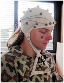
Electrodes are electrical conductors--meaning that electrical charges can travel through them. They are used in scalp EEGs to monitor the electrical activity of the brain, as shown here. They are also used in EKGs (electrocardiograms) to monitor the electrical activity of the heart, and in other monitoring
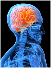
The human body is largely controlled by three pounds of fat and protein--the complex organ that we call the brain. There are some 100 billion neurons in the brain that transmit and receive electrical signals related to thoughts, feelings, movements, and countless other processes.
Sometimes things go wrong. The disorder epilepsy is characterized by seizures that result from the abnormal, excessive firing of neurons in the brain, which often happens for no apparent cause. In cases where medicine is not effective at controlling the seizures, some patients benefit from a surgery that involves removing or isolating the part of the brain causing the seizures. However, this requires knowing the exact location of the brain where the seizure-causing activity originates.
A new brain sensor developed by a team of researchers working at the intersection of materials science, neurology, and electrical engineering could represent a significant improvement in the ability to detect exactly where abnormal brain activity starts in a person with epilepsy, and how seizure activity progresses through the brain.
The first step in determining the seizure onset zone, the physical location where seizures originate, is to monitor the patient's brain activity before, during, and after a seizure. This is usually done with techniques like scalp electroencephalograms (EEGs) and functional MRIs, among others. But if these results are inconclusive, doctors may go one step further and place an array of electrodes directly on the patient's brain, for a test called an electrocorticogram (ECoG).
An ECoG is more informative than an EEG because the electrodes, which detect the electrical signals in the brain, are much closer to the brain activity-- the signal doesn't have to be strong enough to travel through the scalp in order to be detected. This means that an ECoG can create a more detailed map of brain activity and better distinguish where a seizure originates.
Most ECoGs use an array of 16 electrodes that are attached to a frame, or a more flexible grid structure that can have up to 64 electrodes spaced about 1cm apart. Although this technique provides a much better picture of brain activity than an EEG, the surface of the brain is far from smooth. You might recall from biology class (or maybe from a gelatin mold or stress ball) that the surface of the brain is very wrinkly, with lots of folds and grooves, so electrodes placed even 1cm apart are still missing out on a lot of the intricacies of the brain.
This is where the new design has potential. The design incorporates something called flexible electronics, which enables the array to conform much more closely to the wrinkly, folded surface of the brain and pack in hundreds of electrodes.
The term flexible electronics refers to mounting electronic components (like transistors, resistors, capacitors, diodes, etc.) on a flexible surface, instead of on a rigid chip. Scientists have been exploring flexible electronics for several years because of the flexibility they provide when space or shape is concerned. They are used, for example, in cell phones, solar cells, keyboards, and even electronic tattoos. A team of researchers with expertise in biology, electrical engineering, materials, and neuroscience joined forces to redesign the array and incorporate flexible electronics technology, with funding from the National Institutes of Health.
| Other Examples of Flexible Electronics | ||
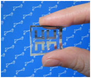 An electronic memory chip created by researchers at NIST (National Institute of Standards and Technology) that can be bent or twisted and still keep functioning. An electronic memory chip created by researchers at NIST (National Institute of Standards and Technology) that can be bent or twisted and still keep functioning.Photo Credit: Electronics and Electrical Engineering Laboratory, NIST |
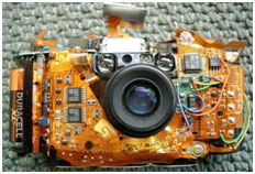 Olympus Stylus camera with skins removed, showing flexible circuit assembly. | 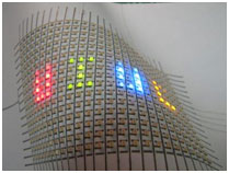 A flexible array of LEDs mounted on paper. Hand-drawn silver ink lines form the interconnects between the LEDs. Photo Credit: Bok Yeop Ahn |
Instead of having wires connected to each of the up to 64 individual electrodes on the array, the new design links the electrodes together electronically within the array, so much less wiring is needed. With the new design, as many as 360 electrodes can fit in the space previously occupied by only 64! The new array can fit right to the contour of the brain and take nearly 300 additional data points, creating a much more detailed map of the brain. Ultimately, this could enable doctors to pinpoint the origin of seizures more accurately and down to a smaller region, possibly reducing the amount of the brain that is removed during surgery.
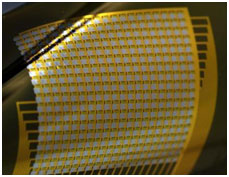
The new electrode array developed by Brian Litt (University of Pennsylvania School of Medicine), Jonathan Viventi (Polytechnic Institute of New York University and New York University). Photo Credit: Travis Ross and Yun Soung Kim, University of Illinois at Urbana-Champaign.
The new design is also significantly thinner--instead of being about as thick as a credit card, the new design is thinner than a human hair. This, combined with the flexibility of the array, means that at some point doctors might be able to drill a hole in the skull and insert a rolled up version of the array into the brain. Then, they could unroll the array once it's inside. This would make the surgery much less invasive since doctors wouldn't need to expose a section of the skull large enough for the entire array.
This new array hasn't been approved for use in humans, but it has undergone preliminary testing on cats, whose brains are physically similar to humans. This small device could have a lot to teach us about the mysteries of the brain, and will hopefully be able to provide some much needed relief to people suffering from epilepsy.
For more information, check out these resources:
Technology Review: An Ultrathin Brain Implant Monitors Seizures
Read more about the new array design, its potential, and what researchers have already learned about the brain activity caused by seizures.
National Geographic: Brain
Explore the anatomy of the brain.
Epilepsy Foundation
Explore resources related to epilepsy and life with epilepsy through these extensive links.
St. Louis Children's Hospital: Subdural Electrode Recording
Read more about how ECoGs are used to help children with epilepsy from St. Louis Children’s Hospital, a pioneer in the technology.
MedlinePlus: EEG
Find out how EEGs are performed, why people have them, and what they look for.


