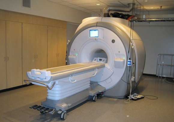
fMRI – The Future Mind Reader?
H. M. Doss
fMRI’s might be the future technology to read your thoughts and emotions. There have been claims that fMRI can determine if you are telling the truth, what image you are looking at, and perhaps - in the future - what you are thinking about, feeling, or your intentions (see these video1, video2).
The physics
Functional Magnetic Resonance Imaging (fMRI) and Magnetic Resonance Imaging (MRI) are a combination of some neat physics and some powerful software that allows soft tissue and blood to be imaged. The physics behind these tools is called nuclear magnetic resonance or NMR. Nuclear Magnetic Resonance was first observed and described in 1938 by Isador Rabi, for which he won the Nobel Prize in physics. It has been studied and used as a technique to determine the structure of compounds for a while.
The processes behind NMR have to do with magnetism and quantum mechanics. Imagine you have a magnetic compass needle that can rotate in all directions. Now imagine putting this magnetic needle in a strong magnetic field. When the magnetic needle and the strong magnetic field interact, the magnetic needle will be forced to line up in a special way with the strong magnetic field. Protons and neutrons, which make up the nucleus of atoms, behave as tiny subatomic magnets due to their quantum property called spin. Spin is a type of angular momentum, but it is not the same as a spinning top. Depending on how the protons and neutrons are placed in the energy levels of a nucleus, and how many protons and neutrons are in the nucleus, determines the overall magnetic moment or “magnetic strength” of the nucleus. Nuclei that have an overall magnetic strength (or spin) behave as subatomic “magnets” that are in constant motion and point in different directions.
When a nucleus with an overall magnetic moment is placed in an external magnetic field it will either line up parallel or antiparallel (often termed spin up or spin down) with the external magnetic field, as shown below. This defines two energy states that are determined by the type of nucleus and the external magnetic field. The difference between these two energy states is proportional to the strength of the external magnetic field that the nucleus is in. As the external field increases, the energy difference between the two states increases. The energy difference is usually related to an amount of energy resonant with a radio frequency pulse. When a radio pulse is applied to the system, it can flip the spin, causing this subatomic magnetic needle to align itself antiparallel to the external magnetic field (the higher energy state).
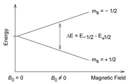
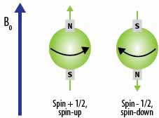
Energy splitting of nuclear spin states as a function of an external magnetic field, B0.ms = +1/2 is the spin up state and ms = -1/2 is the spin down state.
A top or a gyroscope can spin about its own axis and rotate around the axis of the gravitational field it is in. This is called precessing. Just as a top or a gyroscope can precess about the axis of a gravitational field, the nuclear spin states can precess about the axis of the external applied magnetic field. Interestingly, it turns out that the frequency of rotation is equal to the frequency associated with the energy difference between the two spin states. This frequency is called the Larmor frequency. You can learn more about the Larmor frequency at this link. Remember that for most atoms this frequency is in the radio frequency range.

Figure 1 Diagram of Precessing
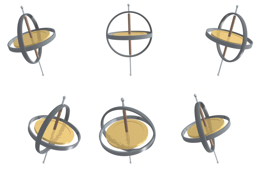
Figure 2 Gyroscope Precessing
If a radiofrequency pulse is emitted of just the right amount of energy for the atom to absorb, the lower energy spin states will flip to the higher energy state. When the atoms go back to the lower energy state, they emit a radio frequency. When many atoms absorb and then re-emit this energy there is a detectable signal.
What does this have to do with “reading” our minds?
For regular MRI’s on our body the hydrogen nuclei in water molecules are used. Our bodies are more than 72% water, which is mainly found in soft tissue. A weighting is often done between the absorbed and emitted radio frequency signals to determine the different types of tissue. The variables a radiologist uses are spin density, two types of relaxation times of the spin state after the applied radio frequency pulse, flow (for example, water through tissue), and spectral shifts (for example, from depths of the tissue). Magnetic fields of up to 3 Teslas (50,000 times the strength of Earth’s magnetic field) are used in MRI machines, which may cost anywhere from half a million to two million dollars. Then, by using some powerful mathematical computations and imaging techniques, the signals observed are transformed into an image of what absorbed and released these signals.
For fMRIs the focus is on blood, blood flow, and how much oxygen is in the blood. During a thought neurons are activated or are being fired in regions of the brain. These active neurons need energy and they get it from glucose. To get the glucose fast there is an increased flow of oxygenated blood to the regions of the brain where the neurons are firing. Oxygen rich blood has about a 20% greater magnetic strength than deoxygenated blood, which was first observed in the 1930’s by Linus Pauling. Because of this difference in magnetic strength, a contrast between the oxygenated and deoxygenated blood can be observed using magnetic resonance imaging techniques. The oxygenated blood produces the strongest signal when placed in a strong magnetic field when just the right radio frequency pulse is applied. Radiologists interpreting fMRI scans measure the blood flow, blood volume, and oxygen levels, which is commonly known as the Blood-Oxygen-Level-Dependent or BOLD signal. A computer processes the detected signal and maps it into 3-D images that are colored based on the measured BOLD signal by using powerful software designed by physicists and medical researchers.
The brain activity is mapped into cubes called voxels, which as similar to computer pixel (picture element). Each voxel (volumetric pixel element) represents tens of thousands of neurons or nerve cells. Most people have the same basic regions of the brain activated when thinking about the same object, when they have visited the same place, and when they perform or think about doing mathematical functions such as addition or subtraction. Radiologists cannot use fMRI to determine each neuron’s function, but they can get a mapping of active parts of the brain and they are getting better at interpreting these maps.
The good news is that fMRI and MRI machines are harmless with virtually no side effects. However they are not for those who have claustrophobia or any metal pins or rods in their bodies. They are effective to determine areas of thought, brain function, and the health of brain tissue, as well as analyzing emotion.
Current Research
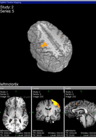
Images of increased neural activity in response to a motor task. Reproduced with permission by Thomas T. Liu, UCSD fMRI Center.
Functional magnetic resonance images (fMRIs) are becoming popular in both the consumer and research arenas. Big businesses use fMRIs to determine consumer preferences and which movie scenes viewers prefer. Smaller businesses utilize the machines as lie detector tests. However, the technique of fMRI is being most utilized for clinical and basic research by many different professions.
The fMRI Center at the University of California at San Diego has about fifty projects going on at the facility ranging from clinical studies to basic research. Some clinical studies look into the causes of Alzheimers, schizophrenia, Parkinsons, autism, alcoholism, and eating disorders. Some basic research tries to answer questions such as: How learning and decision making are affected by drug abuse? How does the brain stop or inhibit an action? How do you learn without instructions? How does vision affect attention? What are some affects of smells, tastes, and language processing?
Many different methods are used in these studies. Some methods map the wiring of the brain and see how the connections of different pathways are affected by different stimuli such as caffeine. Studies show that caffeine reduces connectivity involved in newly learned motor skills, and napping increases connectivity in the brain. It is believed that the brain learns during the resting period by practicing newly learned tasks.
It has also been found that when people are given biofeedback such as a graph of the active area of their brain they can control their brain activity. People are capable of reducing the level of brain activity based purely on the biofeedback, without knowing why that part of the brain is activated.
Scientists are trying to find out what is common across measuring devices to learn how different parts of the brain talk to each other. Mapping the brain is similar to mapping the human genome. Most people use similar parts of the brain for similar tasks, however other parts of the brain are also functioning that are triggered by personal experiences involving a task. Imaging brain activity is just another source of data that provides insights into individual characteristics.
For more about the research going on visit: UCSD fMRI
Links/Reference
1. http://en.wikipedia.org/wiki/Nuclear_magnetic_resonance
2. The Basics of NMR, Joseph P. Hornak,Copyright © 1997-2010 J.P. Hornak.
3. Nuclear Magnetic Resonance Spectroscopy
4. Saenz, A. fMRI Reads the Images in Your Brain – We Know What You’re Looking At, Singularity Hub (17 March 2010)
5. Kay, K.N., Naselaris, T., Prenger, R.J., & Gallant, J.L., Identifying natural images from human brain activity, Nature 452, pp. 352-355 (20 March 2008)
6. Naselaris, T., Prenger, R.J., Kay, K.N., Michael Oliver, M. & Gallant, J.L., Bayesian Reconstruction of Natural Images from Human Brain Activity, Neuron, 63, 6, pp. 902-915, (24 September 2009)
8. About functional (MRI), This website, created by Columbia Medical University, has links to current media discussions and research in the area.
9. Mind Reading =-fMRI – Machine that Reads Your Thoughts – 60 minutes


