Do You See What Eye See?
It’s been hard to miss the publicity for LASIK, the laser surgery that reshapes the cornea to improve the eye’s ability to focus. Actually, both the cornea and the lens focus light, as shown in the diagram. But the lens, itself mostly water, is bathed in watery fluid on both sides, so upon entering and leaving the lens, light bends, or refracts, relatively little. It refracts far more when passing from air into the cornea.
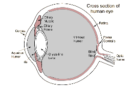
The structure of the human eye. Note that the lens is bathed in watery fluid on both sides, so its refracting power is much less than that of the cornea. (image courtesy of HyperPhysics, by Rod Nave, Georgia State University).

To demonstrate the precision of ablation of human tissue, IBM scientists cut these slots in a human hair with the excimer laser (image courtesy of IBM research).
Reshaping the cornea can make a big difference. Six months after LASIK surgery, about 95% of patients have uncorrected vision of at least 20/40, the minimum for driving, and about 50% have uncorrected vision of at least 20/20, considered to be ideal normal vision. On the downside, about 5% of patients experience side-effects and 1% suffer serious, vision-threatening problems.
LASIK is performed with an ultraviolet (UV) excimer laser. Excimer stands for excited dimer, an excited, unstable molecule of an “inert” gas and a halogen—argon and fluorine. This short-lived molecule dissociates promptly with the emission of a UV photon of a particular frequency. In an alternate process, if a photon of this frequency hits an as-yet-undissociated dimer, the dimer emits a second photon, in step with the first—a process called “stimulated emission,” the basis of laser action. With the argon and fluorine confined in a tube capped with mirrors, one of which allows some light to escape (see diagram), the result is an intense UV laser beam.
Excimer lasers, unlike the familiar ones in bar-code readers, are pulsed—they pack their output into short bursts about 10 nanoseconds long (10-8 sec). This pulsing makes it ideal for eye surgery, because the intense pulses vaporize tissues without heating the rest of the eye. The UV light is absorbed in a very thin layer of tissue, decomposing that tissue into a vapor of small molecules, which fly away from the surface in a tiny plume. This happens so fast that nearly all of the deposited heat energy is carried away in the plume, leaving too little energy behind to damage the adjacent tissue. The process is called ablation, and its application to surgery was an invention of IBM physical scientists. To see its precision, look at the image of the slots cut by an excimer laser in a human hair. Subsequently, the IBM scientists collaborated with ophthalmologists, giving birth to laser refractive surgery.
Ablation of the outermost layers of the cornea and its covering can produce a number of visual problems after the surgery. To avoid this, the ophthalmologist first shaves a thin slice of the outer corneal tissue, folds it back (see photo), and then with the laser ablates the underlying cornea to produce the required shape. When the flap is folded back in place—no sutures are necessary—the two corneal surfaces grow together and the eye usually heals within a week or less. The drawback of this procedure is that cutting the flap is responsible for most of the side-effects.
To reduce these problems, an interdisclipinary team at the University of Michigan is working with the femtosecond laser, whose pulses last only 10-13 seconds.
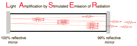
Schematic diagram of laser action. Each wiggly line represents a photon. Note how the number of photons increases through stimulated emission along the path of the original photon. (image courtesy of HyperPhysics, by Rod Nave, Georgia State University).
Research
In the original LASIK, the corneal flap is cut with a microkeratome, a device similar to a carpenter’s plane. Any metallic shards or irregularities in the blade can easily damage the delicate cornea. To avoid this problem, a team of physicists, engineers, and ophthalmologists at the University of Michigan developed a procedure to make this cut with the intense pulses of the femtosecond laser.
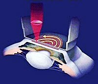
To avoid the side-effects associated with a cut made by a microkeratome blade, the ophthamologist cuts the flap with the laser itself (image courtesy of Intralase).
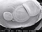
Flap (right) and disk of corneal material (left) removed from a pig’s eye in a procedure performed entirely with an excimer laser (image courtesy of Center for Ultrafast Optical Science, University of Michigan. http://www.eecs.umich.edu/USL/).
The pulses of the femtosecond laser last only 10-13 seconds, as opposed to 10-8 seconds for the excimer, and these ultrashort pulses are made much more intense by a technique called chirped pulse amplification. In general, a laser pulse is amplified by passing it through additional matched lasers, where stimulated emission can vastly increase the number of photons. At very high power, the amplified pulse can have so much energy that it could destroy these lasers. To sidestep this effect, a group at Michigan’s Center for Ultrafast Optical Science developed a way to spread out the different frequencies in the pulse with diffraction gratings to produce a much longer and less intense pulse that can be amplified, as shown in the drawing. Then, with amplification complete, the femtosecond pulse is reconstituted.
Getting back to LASIK, to avoid the damage caused by the microtome, surgeons cut out the underside of the flap with femtosecond laser pulses. These pulses are focused inside the cornea and vaporize tissue at the focal point. The result is a short-lived bubble of gas, which dissolves into the water in the cornea. A rapid sweep of the focus creates a surface of bubbles that define the underside of the flap, and a second cut, which is cylindrical, enables the surgeon to fold back the flap. At this point, the excimer laser performs the LASIK surgery, essentially as before.
Beyond cutting the flap, the Michigan team is developing a way to perform LASIK entirely with the femtosecond laser. In this surgery, the laser ablates two curved surfaces, which define the material to be removed. The upper surface is also the underside of the flap. The surgeon then folds back the flap and removes the “lenticle” of corneal tissue underneath. The image shows the flap, and the disk-shaped tissue that was removed, when this experimental procedure was performed on a pig’s eye.
A further possible procedure avoids the flap altogether. Here the laser would ablate a lenticle-shaped tissue within the cornea, the bubbles would be absorbed in the watery tissue, and the upper and lower surfaces of the ablated lenticle would grow together, providing a reshaped cornea.
This kind of research, not to mention the development of LASIK itself, is made possible by collaboration among physicists, engineers, and MDs. The story is the same for numerous other advances in high-tech medicine, such as the PET scan, the pacemaker/defibrillator, and the endoscope, to name but a few. The corresponding fields of physics range from elementary particles to electricity and magnetism to optics, and more.
Links
U. of Michigan, Center for Ultrafast Optical ScienceHyperPhysics, Georgia State University
Bell Labs
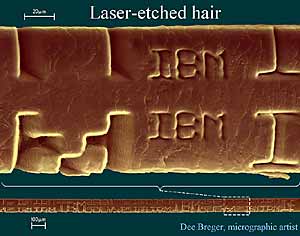
A strand of hair etched with pulses from a laser. Those with access to the device preferred it 3 to 1 to the old method of splitting hairs manually. (Image courtesy of IBM Research)


