MRI Magic
Not an X-Ray
Medical x-rays provide images of the body but utilize radiation that in large doses can damage cells. A completely different technology, magnetic resonance imaging (MRI), emerged in the late 1970s. It produces highly detailed images of soft tissues and requires no x-radiation. MRI is based on nuclear magnetic resonance, a technique developed to probe molecular structure. Ironically, public anxiety about radioactivity and nuclear energy kept the word “nuclear” out of the name of this new kind of imaging, which otherwise might have been called NMRI (nuclear magnetic resonance imaging).
Precessing Protons
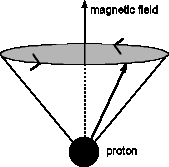
Proton in a magnetic field: The arrow from the proton shows the proton’s spin axis; the tip of this arrow moves in a circle perpendicular to the magnetic field.
To understand the MRI, let’s think about a molecule that contains hydrogen, like a molecule of water or fat. The nuclei of these hydrogen atoms are protons. Each proton possesses an intrinsic spin, so that it acts much like a spinning top. Now suppose we put the molecule between the pole-pieces of a magnet, so the protons are in a magnetic field. Each proton now turns around the field direction like a little gyroscope. (See diagram.) This motion is called precession.
As they precess, the protons, according to quantum mechanics, can have only two orientations. Because of the magnetic field, these two orientations correspond to slightly different energies. If we shine radio waves on these protons, and the radio waves have just the right frequency, the lower energy protons can absorb photons of radiation and flip over. (Quantum mechanics tells us that the photon’s energy is proportional to its frequency.) By observing the details of this energy absorption, which depend on the environment of the protons, we can figure out, for example, if the protons are in water or in fat.
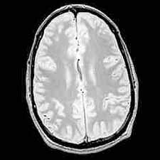
If we analyze many such measurements, we can get an image of a “slice” of the body, as shown. Notice the detail can be seen in the soft tissues. MRI images can show features as small as 1 mm.
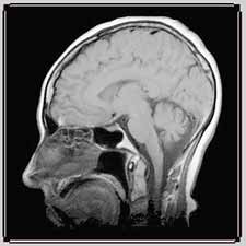
Left: MRI image showing a vertical section of the brain (© 2000 Joseph Hornak, The Basics of MRI)
Right: MRI image showing a horizontal section of the brain (© 2000 Joseph Hornak, The Basics of MRI)
Resonance
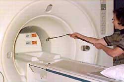
Magnetic force on belt buckle from MRI magnetic field levitates belt. (© 2000 Joseph Hornak, The Basics of MRI)
Resonance is a general phenomenon that is quite important. A periodic system, like a playground swing, repeats its motion or pattern regularly as time passes. The frequency of the system is the number of repeats per second. If someone pushes a swinging child in synch with the swing’s motion, pushing when the swing is already moving away, the push adds energy to the system and the child swings higher. If the frequency of the push equals the frequency of the swing, the maximum amount of energy is added to the system per push. The swinging and the pushing are in resonance with each other. Similarly, in MRI, if the photon has just the right frequency, its energy equals the difference between the energies of the protons’ two orientations, and the collection of photons will absorb the maximum amount of energy.
Your MRI
If you have an MRI, you will recline with part or all of your body inside a large housing, about two feet in diameter. This housing contains a superconducting magnet, which will create a large magnetic field (See Physics in Action Archives, Superconductors, Superconducting Magnet Applications, MRI Scanners). You will wear a special gown to ensure that no magnetic materials are brought into the magnet. The photo of the levitating belt-buckle shows the strength of this magnet.
The exam lasts between a half an hour and an hour. You will have to maintain the same position for minutes at a time. A radiologist, a medical doctor trained to read images, will interpret the results.
Research
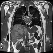
In the quarter century since MRI systems were invented, MRI imaging has steadily improved. Better magnets have led to large bore MRI machines with more spacious housings. The machines can accommodate larger patients and are less likely to frighten people who dislike the cramped spaces of conventional MRI machines. Improved electronics and signal processing systems have increased the detail visible in MRI images and even allowed researchers to collect microscopic images of insects and other tiny samples. MRI images also make up a substantial portion of the National Library of Medicine's Visible Human Project, which is a compilation of detailed layer-by-layer images of a pair of complete male and female human bodies.
Recently, MRI machines have been developed to image and track physiological changes in images of humans and animals as they occur from moment to moment. MRI-based functional imaging non-invasively maps subtle increases in blood flow and oxygenation corresponding to changes in brain activity. Functional MRI (fMRI) has revolutionized research in human brain function and is making headway in many clinical applications.
In effect, fMRI provides movies of the brain at work as neurons become more active in response to a stimulus, mental activity, or mental state. Regions associated with basic sensory and motor activity have been studied, as have regions associated with thought processes and changes in mood or attentiveness. Also, specific clinical populations have shown significant differences in fMRI - based brain activation maps, suggesting that the technique can be used towards more accurate diagnoses and assessment of therapy.
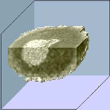
Left: A microscopic MRI image shows surprising detail in a Xenopus oocyte (a frog egg cell), including structure in the light gray cell nucleus. (Image courtesy of the Pacific Northwest National Laboratory, Biomolecular Systems Initiative).
Right: This MRI cross section of an abdomen is one of thousands of images in the The Visible Human Project database that provide a highly detailed survey of the human body.


