CT Scans
Traditional X-Ray Images
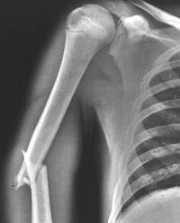
William Roentgen made the first x-ray image in 1895, but the technology remained essentially the same until the late 1960s. These images were projected onto flat detectors, such as film or electronic sensors. The x-rays pass through the body and project a shadow of the contents of the body onto the detectors, which record this projection in various shades of gray. The traditional x-ray image of a broken arm shows that bone, which stops a lot of x-rays, appears white. Soft-tissue, which does not stop x-rays as well as bone, appears a darker gray. The air around the patient, which stops hardly any x-rays, appears black.
CT Scan
Although bones show up clearly on such x-ray images, soft tissues do not show up as well. Moreover, since three-dimensional body parts are projected onto two-dimensional film, much information is lost. What is needed is a “cross-section” view that displays a thin slab of the body. Computed Tomography (CT) images provide this kind of view, as shown in the drawing. A CT image is composed of pixels, whose brightness corresponds to the absorption of x-rays in a thin rectangular slab of the cross-section, which is called a “voxel.”
Notice that the x-ray tube and detectors rotate around the patient. For each direction of the x-ray beam, the scanner records the x-ray absorption by the patient's body. A computer program then computes the brightness of each pixel from all of these separate recordings. Since the CT scan requires so many x-ray exposures, the amount of radiation used to make a CT scan is typically greater that used to make a traditional x-ray.
Introduced in the early 1970s, CT scanning gained rapid acceptance in clinics and hospitals. A physicist and an engineer, Allan Cormack and Godfrey Hounsfield, shared the Nobel Prize in Physiology or Medicine in 1979 for their contributions to its development.
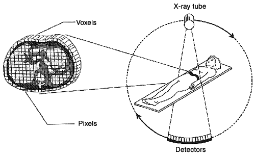
The geometry of the CT Scan; the x-ray tube and detectors rotate, with the axis of rotation running from the patient's head to toe. (image courtesy of the Mayo Clinic)
Improvements in CT Technology
As shown in the table, the technology of CT scanners has improved dramatically since the first scanner was introduced. Today's scanners can image the entire abdomen and pelvis of most adults, making a total of 80 CT images, in less than 30 seconds. The amount of detail in the image has increased six-fold since 1970.
| Specifications | First CT Scanner (circa 1970) | State of the Art CT Scanner (2001) |
| Time to acquire one CT image | 5 minutes | 0.5 seconds |
| Pixel size | 3 mm x 3 mm | 0.5 mm x 0.5 mm |
| Number of pixels in an image | 6,400 | 256,000 |
This pair of CT images of the human brain shows how much image quality has progressed over the last 27 years.
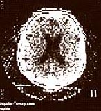
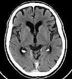
Left: Circa 1975, in the early days of the CT scan.
Right: A present-day scan, showing the six-fold increase in detail
(images courtesy Siemens Medical Systems and Imaginis.com)
Here is a cross-sectional image of the abdomen made with present-day technology.
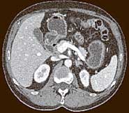
This CT image shows a cross-section of the abdomen. The darker tissue surrounding the abdomen corresponds to a thin layer of fat, and the skin is the very thin white edge of the image. The white, bony structures correspond to the spine (at the bottom center) and short cross-sections of ribs. Blood vessels can be seen at the upper right. (image courtesy of the Mayo Clinic)
Research
Research teams in CT imaging typically include radiologists, physicists, image scientists, computer programmers, engineers, and technicians. Frequently teams work in collaboration with the manufacturers of the CT systems. Preliminary work is commonly performed on inanimate test objects called phantoms and only then does the team performs studies on live patients. Once the results are established, the research team shares its findings in scientific meetings and journal articles.
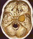
A 3-dimensional (3-D) image of blood vessels within the brain. The large round object is an aneurysm, a ballooning of a blood vessel. The soft-tissue brain matter is not shown in this image. (image courtesy of the Mayo Clinic)
These rapid advances in technology are enabling dozens on new clinical applications, including:
- CT imaging of the heart and coronary arteries to screen for signs of early heart disease
- Emergency room CT scanning procedures to quickly image the entire body of trauma patients.
- Blood flow imaging in the brain to diagnose stroke
- 3-D and virtual-reality imaging of the interior of the colon and lung
- Sophisticated 3-D volume rendering of the bones and soft-tissue organs
The 3-D images mentioned above are produced by computer processing that shows human anatomy in a remarkable new way, by stacking together many cross-sectional images.
Medical Physics
The application of physics to medical imaging is a part of the field of medical physics. Medical physicists work closely with medical doctors and are found in universities, medical schools, and medical research institutes, as well as community hospitals and clinics. They often specialize in one of three main areas:
- Medical imaging (including MRI , nuclear medicine and ultrasound imaging as well as x-ray and CT)
- Radiation oncology (treatment of cancer with various kinds of radiation)
- Medical Health Physics (protection of workers and patients from radiation - both ionizing radiation, like x-rays, and non-ionizing radiation, like lasers and magnetic fields)
Typical Professional Training for Medical Physics:
- B.S. in physics or closely-related field
- M.S. or Ph.D. in Medical Physics
- One or two years of clinical or hospital training
The Physics Central team wishes to express special thanks to Cynthia McCollough of the Mayo Clinic for her help in preparing this feature. More information regarding medical physicists may be found at the American Association of Physicists in Medicine.
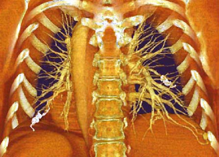
This is a 3-dimensional image of a CT scan of a human chest. Computer processing has removed the front and back of the ribs to reveal bony anatomy (ribs and spine, in white) as well as soft tissue (in red) and blood vessels of the lung (yellow). The carrot-shaped feature is the aorta, the large artery that carries blood from the heart to the body. (image courtesy of the Mayo Clinic)
Links
Physics 2000
PBS
Imaginis
University of Texas
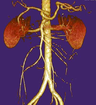
This 3-dimensonal (3-D) image shows the kidneys (in red) and the important blood vessels (in yellow) that supply blood to the kidneys. This type of image is used by surgeons to assess the location of blood vessels prior to a kidney transplant. (image courtesy of the Mayo Clinic)


