Virus Microscopy

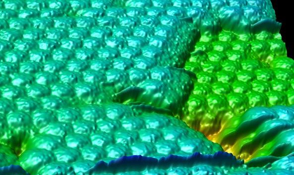
An atomic force microscopy (AFM) scan reveals several hundred tobacco mosaic virus particles.
Image Credit: Alex McPherson/Univ. of Calif. Irvine
Atomic force microscopes detect minuscule changes in force between a probe tip and a sample to produce images of tiny objects. The microscope's probe is attached to a small cantilever, and the cantilever's angle changes as the sample's topography changes. Scientists reflect a laser beam off of the cantilever and detect the subtle changes in the deflected light as the cantilever's angle adjusts to the changing surface of the sample.
More Information:


